Loculated Pleural Effusion Cxr - Health CXR - Zebra Medical Vision | Medical Imaging & AI - Pf ada levels, nodular lung lesions, and loculated pleural effusion may help differentiate tpe from ppe in patients with pf showing.
Loculated Pleural Effusion Cxr - Health CXR - Zebra Medical Vision | Medical Imaging & AI - Pf ada levels, nodular lung lesions, and loculated pleural effusion may help differentiate tpe from ppe in patients with pf showing.. Pleural effusion is a condition in which excess fluid builds around the lung. Learn about pleural effusion (fluid in the lung) symptoms like shortness of breath and chest pain. What does pleural effusion mean? A pleural effusion is accumulation of excessive fluid in the pleural space, the potential space that surrounds each lung. Pleural fluid/serum protein ratio >0.5.
There is a large left pleural effusion obscuring the lower half of the left hemi thorax. Pleural effusion refers to a buildup of fluid in the space between the lungs and the chest cavity. Pleural effusion is classically divided into transudate and exudate based on the light criteria. Pleural fluid/serum protein ratio >0.5. Among the causes, pleural infection, heart failure, and malignan.
CREST-синдром фото from images.radiopaedia.org Pleural fluid ldh > two thirds of upper limit for serum ldh. If none is present the fluid is virtually always a transudate. Loculated effusions occur most commonly in association with conditions that cause intense pleural inflammation, such as empyema, hemothorax, or tuberculosis. Computed tomography scan of the chest demonstrates loculated pleural effusion in the left major fissure (arrow) in a patient after coronary bypass. Pleural effusion occurs when too much fluid collects in the pleural space (the space between the two layers of the pleura). Pleura inflammation, causing sharp pain with breathing; In healthy lungs, these membranes ensure that a small amount of liquid is present between the lungs. Commonly from congestive heart failure or malignancy.
Commonly from congestive heart failure or malignancy.
Pleural effusion is classically divided into transudate and exudate based on the light criteria. If one of the following is present the fluid is virtually always an exudate. Always do pleural biopsy if you suspect tb.disorder in the workup of a pleural effusion after performing thoracentesis always order. Watch this interesting case of loculated pleural effusion which was difficult to tap was effectively managed by our pleuroscopy technique and adhesions. Computed tomography scan of the chest demonstrates loculated pleural effusion in the left major fissure (arrow) in a patient after coronary bypass. Pleural fluid ldh > two thirds of upper limit for serum ldh. Pf ada levels, nodular lung lesions, and loculated pleural effusion may help differentiate tpe from ppe in patients with pf showing. More than one half of these massive pleural effusions are caused by malignancy; Pleural effusion symptoms include shortness of breath or trouble breathing, chest pain, cough, fever, or chills. Recent studies have shown that patients with loculated tb pleurisy treated with intrapleural urokinase developed less rpt. Approximately 1 million people develop this abnormality each year in the united states. Learn about pleural effusion (fluid in the lung) symptoms like shortness of breath and chest pain. Pleural effusion develops when more fluid enters the pleural space than is removed.
It detects pleural effusions with higher sensitivity and specificity than cxr, and provides valuable information about the size and depth of the pleural effusion, the echogenicity of the fluid, the presence of septated or loculated fluid, pleural thickening and nodularity, and the presence of any. Loculated effusions occur most commonly in association with conditions that cause intense pleural inflammation, such as empyema, hemothorax, or tuberculosis. Not respond to chest tube and antibiotics. When you have a pleural effusion, fluid builds up in the space between the layers of your pleura. Pleural effusions occur as a result of increased fluid formation and/or reduced fluid resorption.
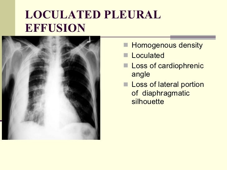
Chest x ray pathology from image.slidesharecdn.com How is pleural effusion detected. Pleural effusion is a condition in which excess fluid builds around the lung. Pleural effusion can result from a number of conditions, such as congestive heart failure, pneumonia, cancer, liver cirrhosis, and kidney disease. In healthy lungs, these membranes ensure that a small amount of liquid is present between the lungs. Not respond to chest tube and antibiotics. Loculated pleural effusion on cxr. Loculated effusion (atypical radiological findings). Commonly from congestive heart failure or malignancy.
Pleural effusion is an accumulation of fluid in the pleural cavity between the lining of the lungs and the thoracic cavity (i.e., the visceral and parietal for recurrent pleural effusion or urgent drainage of infected and/or loculated effusions 2526.
Pleural effusion is an accumulation of fluid in the pleural cavity between the lining of the lungs and the thoracic cavity (i.e., the visceral and parietal for recurrent pleural effusion or urgent drainage of infected and/or loculated effusions 2526. How is pleural effusion detected. What does pleural effusion mean? Pf ada levels, nodular lung lesions, and loculated pleural effusion may help differentiate tpe from ppe in patients with pf showing. Not respond to chest tube and antibiotics. Excess fluid in the pleural space; The lungs and the chest cavity both have a lining that consists of pleura, which is a thin membrane. If none is present the fluid is virtually always a transudate. Pleural effusions occur as a result of increased fluid formation and/or reduced fluid resorption. A pleural effusion is accumulation of excessive fluid in the pleural space, the potential space that surrounds each lung. Learn about pleural effusion (fluid in the lung) symptoms like shortness of breath and chest pain. Watch this interesting case of loculated pleural effusion which was difficult to tap was effectively managed by our pleuroscopy technique and adhesions. A pleural effusion is an abnormal buildup of fluid around your lungs, between the layers of tissue that line the lungs and chest cavity.
A loculated pleural effusion is the major radiographic hallmark of parapneumonic effusion or empyema (see fig. Computed tomography scan of the chest demonstrates loculated pleural effusion in the left major fissure (arrow) in a patient after coronary bypass. Pleura inflammation, causing sharp pain with breathing; Other causes are complicated parapneumonic effusion. The pleura are thin membranes that line the lungs and the inside of the chest cavity and act to lubricate and facilitate breathing.
The patient's chest radiograph (CXR)-large-sized, right ... from www.researchgate.net A loculated pleural effusion is the major radiographic hallmark of parapneumonic effusion or empyema (see fig. If none is present the fluid is virtually always a transudate. A pleural effusion is accumulation of excessive fluid in the pleural space, the potential space that surrounds each lung. Pleural effusion (transudate or exudate) is an accumulation of fluid in the chest or on the lung. Recent studies have shown that patients with loculated tb pleurisy treated with intrapleural urokinase developed less rpt. Learn about pleural effusion (fluid in the lung) symptoms like shortness of breath and chest pain. Always do pleural biopsy if you suspect tb.disorder in the workup of a pleural effusion after performing thoracentesis always order. When you have a pleural effusion, fluid builds up in the space between the layers of your pleura.
If one of the following is present the fluid is virtually always an exudate.
Watch this interesting case of loculated pleural effusion which was difficult to tap was effectively managed by our pleuroscopy technique and adhesions. Always do pleural biopsy if you suspect tb.disorder in the workup of a pleural effusion after performing thoracentesis always order. Loculated effusion (atypical radiological findings). Not respond to chest tube and antibiotics. Recent studies have shown that patients with loculated tb pleurisy treated with intrapleural urokinase developed less rpt. Approximately 1 million people develop this abnormality each year in the united states. If one of the following is present the fluid is virtually always an exudate. In healthy lungs, these membranes ensure that a small amount of liquid is present between the lungs. Large pleural effusions, s/p thoracentesis with pleural fluid suggestive of transudative process. Pleural effusion occurs when too much fluid collects in the pleural space (the space between the two layers of the pleura). Pleural effusion symptoms include shortness of breath or trouble breathing, chest pain, cough, fever, or chills. The cxr shows classic evidence of congestive heart failure with cardiomegaly, upper lobe venous diversion, and bilateral pleural effusions. Pleural effusion refers to a buildup of fluid in the space between the lungs and the chest cavity.
Meaning of pleural effusion medical term loculated pleural effusion. Pleural fluid/serum protein ratio >0.5.


Posting Komentar
0 Komentar